-
The association
-
Our strengths
-
Our services
-
Training

Analysis under the microscope
Dark field and coagulated blood

Live blood analysis involves taking a single drop of live blood from the fingertip, then placing it under a powerful microscope. The image is then displayed on a screen for both practitioner and patient to view.
Blood carries oxygen, nutrients and other vital agents throughout the body to maintain health.
It is also the means of detoxification, transporting cellular waste to the liver and kidneys for elimination from the body.
Blood can therefore serve as a health predictor, providing an indication of disease long before symptoms appear.
This method of analysis differs from the usual laboratory tests that quantify the levels of certain components in a blood sample. Live blood screening gives an indication of the QUALITY of an individual's blood - an important element of preventive health care that shows the effects that diet, lifestyle and stress can have on our well-being.
Microscopic examination of the shape and function of blood cells and plasma may reveal signs of :
- Poor digestion/diet combination.
- Weakened immune system.
- Vitamin deficiencies
- Intestinal and liver toxicity
- Dehydration
- Stress
- Increased cholesterol and formation of crystals.
- Poor circulation.
- Heavy metal accumulation.
- Presence of parasites / fungi / bacteria.

We use darkfield microscopy as well as brightfield microscopy to perform live and dried blood analysis on a customer, then classify the anomalies observed and decide which problem to focus on based on our analysis.
Live blood analysis is revolutionizing many natural health practices around the world, giving practitioners the edge they need to achieve better results in their practice.
Dark field/blood smear analysis is also known as naturopathic microscopy, live blood cell analysis, nutritional microscopy and oxidative stress testing.
Live blood cell analysis is the observation of live blood cells using a specialized high-power microscope with a camera that projects an image of the live blood cells onto a screen to be viewed by the practitioner and client together, enabling images and videos to be recorded.
A small puncture of blood is placed on a glass slide and visualized on the screen. In live blood analysis, the blood is not dried or stained beforehand, so that the blood elements can be seen in their living state.
The practitioner and consultant examine variations in the size, shape, ratio and fine structure of red blood cells, white blood cells, platelets and other blood structures.
The knowledge gained from dark field/blood smear analysis, correlated with other clinical data, enables the analyst to understand the individual state of health of his clients at a much deeper level.
As a result, an appropriate course of natural treatment and lifestyle/dietary interventions can be formulated and, in addition, the efficacy of various treatment combinations can be tested and progress monitored by observing changes through further analysis of live blood.
Nutritional microscopy uses live blood analysis with a focus on the use of nutrition to naturally achieve optimal health.
Dry blood analysis or oxidative stress testing is another very valuable test in the analysis of living blood cells. With the oxidative stress test (dry blood analysis), we can learn a lot about the level of damage caused by free radicals/oxidative stress and toxins in the body.
All our office consultations offer this service.
NSSR, ADIMOGH and Lyme sessions offer this remote management service.

Online consultations using remote microscopy
ADIMOGH/NSSR/Lyme-Fibromyalgia method


Example of live blood, coagulated blood and blood smear analysis
1. In no case do we seek to diagnose or replace the advice of a treating physician or biologist.
2. These analyses, unlike laboratory analyses, are qualitative and not quantitative.
Here's what you'll find out about our remote cham noir, coagulated blood or smear analyses (from the comfort of your home).
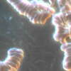
Protein binding
Lemon-shaped chain formations GR.
Generally associated with impaired digestion of dietary proteins, due to excessive protein intake or low production of proteolytic enzymes by the pancreas. Particularly associated with difficulty in digesting dense animal proteins (such as meat and chicken).

Anisocytosis
Red blood cells of variable size, some larger and some smaller than normal.
Most often due to vitamin B12 and/or folic acid deficiency, iron deficiency, trace element deficiency and, in some cases, certain pathological conditions and hereditary forms of anemia.
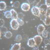
Oxidative stress and inflammation
These forms indicate the presence of inflammation, antioxidant deficiency and pathogen-mediated chronic inflammation. A veritable vicious circle of inflammation and infection.
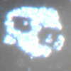
Eosinophilia
Elevated eosinophil counts indicate an imbalanced immune response to parasites, allergens, histamine, etc.
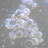
Mast cell activation-SAMA
In consultants suffering from mast cell activation (SAMA), it's common to see an unusual activation of inflammation, a lack of antioxidants, an excess of histamine granulocytes among other manifestations and disturbances.
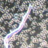
Mycoses and
co-infections
Mycoses and co-infections of the digestive (not respiratory!) apergillosis family can be seen under the microscope. Treatment options
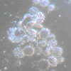
Neutrophilic - Disturbed
Disturbed neutrophils are most often indicative of chronic stress on the immune system, parasitism of white blood cells and an acidic, unbalanced terrain.
They can also be associated with severe toxicity and allergic reactions.
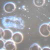
Neutrophils -
Non-viable
Observation of neutrophils is indeed crucial for assessing the state of the immune system. Neutrophils should be actively moving. Round, symmetrical and immobile neutrophils are not viable and suggest an underactive immune system. It can be caused by: mineral deficiencies, ongoing infections and antibiotics, smoking, alcohol, medication and sugar consumption and digestive weakness. Stress, lack of exercise, poor sleeping habits and yeast overgrowth can also contribute.

Intestinal diagram
The observation of a cluster of round white holes in the center of the sample may indeed indicate intestinal challenges.
These challenges may include intestinal inflammation (colitis, enteritis), leaky gut syndrome, strictures, diverticula, irritable bowel syndrome and poor tissue integrity. The presence of intestinal patterns in more than 3 layers indicates that digestive system support is a high priority.

L-form bacteria Lyme family
Unstable bacteria with no cell membrane. Gram-negative or gram-positive, often belonging to the Lyme family, among others.
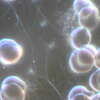
Biofilms
A biofilm is a more or less complex, often symbiotic, multicellular community of micro-organisms, adhering to each other and to a surface, marked by the secretion of an adhesive and protective matrix. This matrix acts as a "mask", preventing allopathic and natural treatments from working.
Bacteria of the Lyme family
Co-infections, babesiosis, bartonellosis, parasitosis and other infections co-existing in people with Lyme can be seen under microscopy.

Online consultations using remote microscopy
ADIMOGH/NSSR/Lyme-Fibromyalgia method
Association Humankind Wellbeing SIRET : 923 516 587 00014 . Organisme de Formation (OF) registered under the activity number 84730284273.
Humankind Wellbeing SAS SIRET 94311513900017


